Glenohumeral joint: Between the humeral head (ball) and the glenoid (socket of the shoulder blade).
Acromioclavicular (AC) joint: Between the acromion (part of the scapula) and the clavicle.
Age-related degeneration: Most common in individuals over 50 years old.
Previous injury: A history of shoulder dislocations, fractures, or rotator cuff injuries can predispose to arthritis.
Overuse or repetitive stress: Repeated overhead movements or heavy labor can accelerate joint wear.
Genetics: Family history of arthritis may increase the risk.
Rheumatoid arthritis: An autoimmune condition that can also lead to shoulder arthritis.
Pain: A deep, aching pain in the shoulder, often worse with movement or at night.
Stiffness: Difficulty moving the shoulder, especially in the morning or after periods of inactivity.
Grinding or crepitus: A grating sensation or sound during movement due to roughened joint surfaces.
Loss of range of motion: Difficulty with overhead activities, reaching behind the back, or rotating the arm.
Weakness: Decreased strength due to pain and muscle atrophy.
Physical examination: Testing shoulder strength, range of motion, and evaluating for signs of rotator cuff dysfunction.
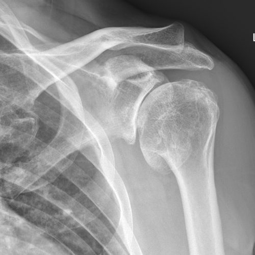
X-rays: Show upward migration of the humeral head, joint space narrowing, bone spurs, and other degenerative changes.
MRI: Assesses the extent of rotator cuff damage, tendon quality, and joint degeneration.
CT scan: Can provide detailed information about bone structure and joint erosion.
Rest: Avoid activities that worsen symptoms.
NSAIDs: To relieve pain and inflammation.
Physical therapy: Focus on stretching, strengthening, and maintaining flexibility.
Corticosteroid injections: Injections into the joint to reduce inflammation and pain.
Hyaluronic acid injections: Lubricating injections to improve joint movement.
Arthroscopic debridement: Cleaning the joint of loose cartilage or bone spurs. This may improve symptoms in the short term but does not eliminate the degenerative progress of arthritis.
Total shoulder replacement (Arthroplasty): Replacing the damaged joint with an artificial implant. This may be done with either an anatomic (standard) or reverse configuration, depending on other patient factors, however, the anatomic total shoulder requires a functional rotator cuff.
Hemiarthroplasty or humeral head resurfacing: Replacing only the humeral head with a prosthesis. Traditionally also used in fracture setting, and largely replaced by total shoulder replacement techniques.
Strengthen the rotator cuff: Proper exercise to maintain shoulder stability.
Avoid overuse: Reduce repetitive overhead activities.
Manage injuries early: Prompt treatment of rotator cuff tears may prevent the development of cuff tear arthropathy.
Regular check-ups: For individuals with prior shoulder injuries or a family history of arthritis.
Massive rotator cuff tears: Tears involving two or more rotator cuff tendons, especially if the tear is chronic or not repairable.
Altered joint mechanics: Without the support of a functional rotator cuff, the humeral head can move abnormally, causing wear and degeneration of the joint.
Age: More common in older individuals due to degenerative changes.
Previous injury: Chronic or untreated rotator cuff tears can lead to cuff tear arthropathy.
Pain: Typically located in the top or outer part of the shoulder. Can be severe and constant.
Limited range of motion: Difficulty lifting the arm, especially overhead. “Pseudoparalysis” can occur, where the shoulder appears paralyzed due to pain and weakness.
Weakness: Significant weakness, especially with lifting or rotating the arm.
Crepitus: Grinding or popping sensation with shoulder movement due to altered mechanics and joint degeneration.
Visible deformity: In severe cases, a high-riding humeral head can be observed.
Physical examination: Testing shoulder strength, range of motion, and evaluating for signs of rotator cuff dysfunction.
X-rays: Show upward migration of the humeral head, joint space narrowing, bone spurs, and other degenerative changes.
MRI: Assesses the extent of rotator cuff damage, tendon quality, and joint degeneration.
CT scan: Can provide detailed information about bone structure and joint erosion.
Rest and activity modification: Avoid activities that exacerbate symptoms.
NSAIDs: For pain and inflammation relief.
Physical therapy: To maintain mobility, flexibility, and strengthen the remaining functional muscles.
Steroid injections: To reduce inflammation and pain temporarily.
Reverse Shoulder Arthroplasty (RSA): A type of shoulder replacement specifically designed for patients with cuff tear arthropathy. In RSA, the anatomy of the shoulder is “reversed” by placing the ball component on the scapula and the socket on the humeral side. This design relies on the deltoid muscle instead of the rotator cuff for movement, providing better function for those with non-functional rotator cuffs.
Hemiarthroplasty: In some cases of extreme scapular bone loss, replacing only the humeral head can be considered, but RSA is generally preferred for severe CTA.
Tendon transfer: In younger patients or selected cases, transferring a tendon from another muscle (e.g., the latissimus dorsi or pectoralis major) to replace lost function may be an option.
Strengthen the rotator cuff: Proper exercise to maintain shoulder stability.
Avoid overuse: Reduce repetitive overhead activities.
Manage injuries early: Prompt treatment of rotator cuff tears may prevent the development of cuff tear arthropathy.
Regular check-ups: For individuals with prior shoulder injuries or a family history of arthritis.
Miami Shoulder Institute is dedicated to delivering world-class care with compassion, expertise, and integrity. Your mobility, our mission.